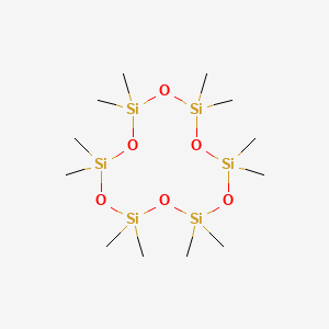

1. 540-97-6
2. Cyclohexasiloxane, Dodecamethyl-
3. Cyclomethicone 6
4. 2,2,4,4,6,6,8,8,10,10,12,12-dodecamethyl-1,3,5,7,9,11-hexaoxa-2,4,6,8,10,12-hexasilacyclododecane
5. Cyclohexasiloxane
6. Xhk3u310ba
7. 2,2,4,4,6,6,8,8,10,10,12,12-dodecamethylcyclohexasiloxane
8. Einecs 208-762-8
9. Unii-xhk3u310ba
10. Hsdb 7723
11. Ec 208-762-8
12. Dodecamethyl Cyclohexasiloxane
13. Schembl93785
14. Xiameter Pmx-0246
15. Cyclohexasiloxane [inci]
16. Dtxsid6027183
17. Iumsdrxlfwagnt-uhfffaoysa-
18. Chebi:191103
19. Cyclomethicone 6 [usp-rs]
20. Mfcd00144215
21. Akos015839990
22. Zinc169794506
23. Fs-5671
24. Dodecamethylcyclohexasiloxane [mi]
25. Dodecamethylcyclohexasiloxane [hsdb]
26. Db-008587
27. D2040
28. Dodecamethylcyclohexasiloxane [who-dd]
29. Ft-0625566
30. S08515
31. T71035
32. Dodecamethylcyclohexasiloxane, Analytical Standard
33. A914553
34. Q27293843
35. 2,2,4,4,6,6,8,8,10,10,12,12-dodecamethylcyclohexasiloxane #
36. Cyclohexasiloxane, 2,2,4,4,6,6,8,8,10,10,12,12-dodecamethyl-
37. 2,2,4,4,6,6,8,8,10,10,12,12-dodecamethylcyclohexasiloxane 95%
38. 2,2,4,4,6,6,8,8,10,10,12,12-dodecamethylcyclohexasiloxane, 95%
39. 2,2,4,4,6,6,8,8,10,10,12,12-dodecamethylcyclohexasiloxane, Aldrichcpr
40. Cyclomethicone 6, United States Pharmacopeia (usp) Reference Standard
41. 2,2,4,4,6,6,8,8,10,10,12,12-dodecamethyl-1,3,5,7,9,11-hexaoxa-2,4,6,8,10,12-hexa
42. D-6
| Molecular Weight | 444.92 g/mol |
|---|---|
| Molecular Formula | C12H36O6Si6 |
| Hydrogen Bond Donor Count | 0 |
| Hydrogen Bond Acceptor Count | 6 |
| Rotatable Bond Count | 0 |
| Exact Mass | 444.11274807 g/mol |
| Monoisotopic Mass | 444.11274807 g/mol |
| Topological Polar Surface Area | 55.4 Ų |
| Heavy Atom Count | 24 |
| Formal Charge | 0 |
| Complexity | 320 |
| Isotope Atom Count | 0 |
| Defined Atom Stereocenter Count | 0 |
| Undefined Atom Stereocenter Count | 0 |
| Defined Bond Stereocenter Count | 0 |
| Undefined Bond Stereocenter Count | 0 |
| Covalently Bonded Unit Count | 1 |
/Researchers/ evaluated the percutaneous absorption of neat D6 radiolabeled with (14)C following application to human skin in vitro. The test substance was applied under semiocclusive conditions in a Teflon flowthrough diffusion cell system. Human epidermis was prepared from abdominal skin (6 donors). The epidermis with the top layer of dermis was separated from the rest of the skin by dermatoming, and the skin samples were mounted in replicate. Skin samples from 3 donors passed the barrier integrity test. A physiological receptor fluid was pumped beneath the skin samples. Skin samples from each of the 6 donors were dosed with neat (14)C-cylohexasiloxane (14C-D6) at a target dose of 6 mg/sq cm during the 24-hour-exposure period. At the conclusion of the assay, the majority of the applied dose was located on the skin surface (46.407% of applied dose) or volatilized from the dosing site and collected in charcoal traps (40.057% of applied dose). Practically no (14)C-D6 penetrated through the skin and into the receptor fluid. The percentage of applied neat (14)C-D6 recovered from all samples that were analyzed was 89.542% +/- 4.154%, which included 3.075% of the applied neat (14)C-D6 (SE of the mean = 0.852% of the applied dose) that was found in the skin. The results of an additional experiment indicated that, after the skin was washed at 24 hours, the portion of (14)C-D6 observed in the skin did not penetrate through the skin, but continued to evaporate. Thus, it was concluded that, under the conditions of this assay, D6 was not percutaneously absorbed.
Johnson W Jr et al; Int J Toxicol 30 (6 Suppl): 149S-227S (2011)
/Researchers/ evaluated the disposition of (14)C-D6 using 10 groups of Fischer 344 rats (CDF(F-344)/ CrlBR strain). The animals were 8 to 10 weeks old and body weight ranges were 163 to 219 g (males) and 133 to 155 g (females). A single oral dose of (14)C-D6 (in corn oil, 1000 mg/kg body weight) was administered to a group of 4 males and 4 females; metabolism cages were used for the collection of urine, feces, and expired air. The animals were killed at 168 hours postdosing, and selected tissues and remaining carcasses collected and analyzed for radioactivity. Expired volatiles and feces were also analyzed for parent D6 concentration. A separate group of rats (6 males and 6 females), cannulated via jugular vein, was used to determine radioactivity and parent D6 concentration in the blood at 15 minutes and at 1, 6, 12, 18, 24, 48, 72, 96, 120, 144, and 168 hours postdosing. Wholebody autoradiography (WBA) was used for qualitative in vivo assessment of tissue distribution of radioactivity in male and female rats after single oral administration of D6 (in corn oil). Animals in the WBA groups were killed at 1, 4, 24, 48, 96, and 168 hours postdosing. In males and females, the majority of the administered dose was excreted in the feces. Based on the recovered radioactivity (urine, expired volatiles, expired CO2, tissues, and carcass), the absorption of D6 was 11.88% (males) and 11.83% (females) of the administered dose. For most of the recovered radioactivity, a similar pattern of distribution of the radioactivity was noted in males and females. However, considerable variability in the levels of radioactivity in expired volatiles was reported, which may have been due to off gassing from the fecal pellets that were not collected, as intended, but remained on the inside of the cage. The authors noted that this phenomenon could potentially produce some false high values for expired volatiles and absorption due to partitioning from the fecal matter into the air. All of the radioactivity in the expired volatiles was attributed to parent D6. Metabolic profile evaluation of the urine and feces indicated that all of the radioactivity in the urine consisted of polar metabolites, whereas, in the feces, the majority was parent D6, with a trace nonpolar metabolite. Whole body autoradiography data supported mass balance data showing that the majority of administered D6 in corn oil stayed in the gastrointestinal (GI) tract and was excreted in the feces within 48 hours. Low levels of radioactivity were detected in organs and tissues, such as the liver, fat tissue, and bone marrow, indicating some absorption of D6. Statistical analysis of blood curves indicated the presence of small amounts of metabolites in the blood, based on the difference between radioactivity and parent AUCs (AUCmetabolites = AUCradioactivity AUCparent).
Johnson W Jr et al; Int J Toxicol 30 (6 Suppl): 149S-227S (2011)
Silicone [poly(dimethylsiloxane)] gel used in breast implants has been known to migrate through intact silicone elastomer shells, resulting in the clinically observable "gel bleed" on the implant surface. Although silicon concentrations in capsular tissues of women with silicone prostheses have been measured with element-specific silicon analyses, no silicone-specific investigation of these tissues has been performed as yet. A combination of element-specific inductively coupled plasma high-resolution isotope dilution mass spectrometry (ICP-HR-IDMS) and species-specific gas chromatography coupled mass spectrometry (GC-MS) was used to analyze silicon, platinum, and siloxanes in prosthesis capsule, muscle, and fat tissues of women (n=3) who had silicone gel-filled breast implants and in breast tissue of non-augmented women (n=3) as controls. In all tissues of augmented women, siloxanes, in particular octamethylcyclotetrasiloxane (D4), decamethylcyclopentasiloxane (D5), and dodecamethylcyclohexasiloxane (D6) were identified. Depending on the siloxane species and type of tissue analyzed, siloxane levels in the range of about 10-1,400 ng/g were detected; total silicon was found in all tissue samples in the range of about 8,900-85,000 ng/g. Higher platinum levels ranging from 25-90 ng/g were detected in fibrin layer and fat tissue of two patients with prostheses. No siloxanes were detected in control breast tissue samples. This investigation of human tissues by a combination of element-specific and species-specific analytical techniques clearly demonstrates for the first time that platinum and siloxanes leak from prostheses and accumulate in their surrounding tissues.
PMID:12589499 Flassbeck D et al; Anal Bioanal Chem 375 (3): 356-62 (2003)
To examine the distribution of low molecular weight silicones in body organs, separate groups of female CD-1 mice were injected with either breast implant distillate composed primarily of hexamethylcyclotrisiloxane, octamethylcyclotetrasiloxane, decamethylcyclopentasiloxane, dodecamethylcyclohexasiloxane, and tetradecamethylcycloheptasiloxane or a polydimethylsiloxane oil containing low molecular weight linear siloxanes. Mice were injected subcutaneously in the suprascapular area and killed at different times. Levels of individual low molecular weight silicones were measured in 10 different organs (brain, heart, kidney, liver, lung, mesenteric lymph nodes, ovaries, spleen, skeletal muscle, and uterus). In mice treated with the cyclosiloxane mixture and killed at 3, 6, or 9 weeks, highest levels of cyclosiloxanes were found in the mesenteric lymph nodes, ovaries, and uterus, but all organs examined contained cyclosiloxanes. In a cohort killed at 1 year, most organs contained measurable cyclosiloxanes with highest levels in mesenteric lymph nodes, abdominal fat, and ovaries. Of the individual cyclosiloxanes measured, selective retention of decamethylcyclopentasiloxane and dodecamethylcyclohexasiloxane relative to octamethylcyclotetrasiloxane was seen in all organs at all time points studied. Organs from animals receiving the linear siloxane mixture were harvested at 9, 12, and 15 weeks. ... Maximum levels in the brain, lungs, and mesenteric lymph nodes /were found/, but all other organs contained measurable levels. These data are, to the best of our knowledge, the first demonstration that after a single subcutaneous injection silicones are widely distributed throughout the body and can persist over an extended period.
PMID:9502404 Full text: https://www.ncbi.nlm.nih.gov/pmc/articles/PMC1858395 Kala SV et al; Am J Pathol 152 (3): 645-9 (1998)
The disposition of (14)C-dodecamethylcyclohexasiloxane (D6) was evaluated in male and female F344 rats following a single oral administration of 1000 mg of (14)C-D6 in corn oil/kg of body weight. Animals (N=4/sex) were housed in glass metabolism cages for collection of excreta and expired volatiles. At 168 hr post-dose, animals were sacrificed and selected tissues and remaining carcasses were collected. All samples were analyzed for radioactivity content. Feces and expired volatiles were also analyzed for parent D6 concentration. A separate group of animals, cannulated via the jugular vein, (N=6/sex) was used to determine radioactivity and parent D6 concentration in blood at predetermined time points. Whole-body autoradiography (WBA) was used for qualitative in vivo assessment of tissue distribution of radioactivity. The majority of administered radioactivity (>80%), regardless of sex, was eliminated in the feces. Total absorption (radioactivity recovered in urine, expired volatiles, expired CO2, tissues and carcass) in males and females was 11.88% and 11.83%, respectively. Both sexes showed similar patterns of disposition (Urine: 0.38% and 0.32%; Expired volatiles: 11.20% and 11.21%; Expired CO2: 0.13% and 0.09%; Tissues: 0.03% and 0.04%; Carcass: 0.14% and 0.17% for males and females, respectively). The entire radioactivity in the expired volatiles was attributed to parent D6. Metabolic profile evaluation showed that the entire radioactivity in the urine consisted of polar metabolites, whereas in the feces the majority was parent D6 with a trace of nonpolar metabolite. The WBA data also showed that the majority of D6 remained in the GI tract and was excreted in feces within 48 hr. Low levels of radioactivity were systemically available and distributed to organs and tissues such as liver, brown fat and bone marrow. Statistical analysis of the blood curves indicated the presence of small levels of metabolites in the blood (approximately 117 ug equivalent D6*hr/g of blood). In summary, approximately 12% of (14)C-D6 delivered in corn oil appeared to be absorbed after a single oral administration to F344 rats.
Plotzke KP et al; Toxicologist 78 (1-S): 23 (2004)
MARKET PLACE


